
Anatomy of Spine: Osteoarthritis |
With a digital imaging, Dr. Jose Mena, Interventional Spine Specialist with Miami Orthopedics & Sports Medicine Institute, explains how the osteoarthritis spine looks.
He describes discs serve as cushion and they’re between the different segments of the spine.
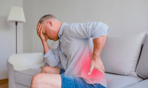
As people age, discs become weaker and joints become arthritic, causing a lot of pain.
Transcript
Right now doctor and we’re looking at spine osteoarthritis doctor that’s correct can you tell us what that is so basically here we have the spine or what I’m seeing is the spine looking from the side view here so basically the belly will be sitting over here okay and the back portion on the back muscle will be down here in this game so basically we have in between in the spine is being formed by the bones on the spine which are this squares that you guys see over here and in between we have this structures which are called the disc the disc they serve as the at the cushion and between the different segments as you can see suppose you can see a normal one how smooth it is the levels and how irregular the levels become so normally what happens as we age the discs they start losing some other water content they will start looking losing some of the the tensile strength they become weaker and so when they become weaker some of the stress normally keeping in mind and they bear about 80% of the load of the spine the res being transferred to those joints that they were referring to in the in the in the clip which are called the facet joints or the sequel of the syncopal CCL joints those joint they then become arthritic instead of being bearing about 20% of the spine at the load is fine now they’re very more more load so we come arthritic they can develop some bone spurs and normally when they develop the bone spur they normally develop over here and as you can see this structure is coming down here is called a nerf so if this starts narrowing they started getting thinner and some of the joints they start in growing over here and causing some some bone spurs the nerve can get compressed and so that’s one of the reason of people developing sciatica and when they get compressed doctor that’s obviously where the pain occurs that’s correct so typically depending on the level some things we might see this on on a normal MRI or I mean on a given an MRI and the patient might be completely asymptomatic so that doesn’t mean because you have those findings on an imaging which we expect as we age that people are going to get the interpreted changes as we age but as just like the gray hair but and some occasions those symptoms those findings can correlate with the symptomatology and that’s basically up to the clinician just to figure it out and address those you