Could my skin lesion be cancerous? |
Skin cancer is the most common form of cancer in the United States, occurring in over two million people each year. Three of the most common types of skin cancer are squamous cell carcinoma, basal cell carcinoma, and melanoma. Each has characteristic features that can help alert you to the possibility that you may have developed a skin cancer.
Squamous cell skin cancers (SCC) arise from squamous cells which comprise most of skin’s outer layer (epidermis). Since these are often related to UV exposure from the sun, they most commonly develop on the face, ears, arms or hands. In their earliest form, they often appear as rough, scaly, red patches. Unlike a skin rash that may fade with time, however, these rough patches persist and continue to progress slowly. Over time, they may become crusted, bleed, or develop an indentation in the center of the lesion. On occasion a SCC may have a wart-like appearance.
Approximately 40 to 60% of squamous cell skin cancers begin as a pre-malignant lesion known as an actinic keratosis (AK). Also known as solar keratosis, these develop on sun-exposed areas of the body such as the face, arms, backs of hands, and lips. An AK is usually rough in texture, resembling a wart, and may be felt before actual changes of the skin are noted. Their color can range from flesh-toned to brown or red. Most are small, less than a half inch across. Approximately 10% of AKs progress to become squamous cell skin cancer.
Basal cell carcinoma (BCC) develops in the basal cell layer of the skin, the deepest layer of the epidermis. It is the most common type of skin cancer occurring in the U.S. BCCs are usually seen in areas of the body that have been exposed to the sun, such as the face or arms. Warning signs that should alert someone to the possibility of a BCC include:
- A persistent open sore that bleeds or oozes. On occasion, these may appear to heal only to re-open and bleed again.
- A pearly bump or nodule that may be pink, red, or white. Small blood vessels may be apparent on the surface.
- A lesion with a crater-like appearance with elevated border and indentation in the center.
- An area of skin that is shiny and tight like a scar that develops without injury.
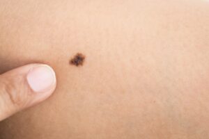
Melanoma is a cancer of the pigment producing cells (melanocytes) of the skin. The great majority of melanomas can be attributed to exposure to ultraviolet (UV) radiation from the sun. Melanoma begins on the surface of the skin but over time can extend deeper and in some cases, even spread throughout the body. Signs indicating that a skin lesion could be a melanoma include new pigmented area on the skin, or a change in size, shape or color of an existing mole. The ABCDE rule is another way to recognize melanomas:
- Asymmetry: A mole that has an irregular shape, or the shape of one half does not match the other half.
- Border: The edges are often irregular, blurred, rough, or notched in outline.
- Color: Most moles are evenly colored, e.g. brown, black, or tan. Changes in the shade or distribution of color throughout the mole can signal melanoma.
- Diameter: Moles larger than ¼ inch (6 mm, the size of a pencil eraser) across are suspicious.
- Evolving: The mole has changed over the past few weeks or months
Regular examination of the skin, by a physician or by self-examination, is the key to identifying skin cancers in their earliest stages. Any suspicious lesion should be brought to the attention of your primary care doctor or a dermatologist. Next article we’ll look at how these common forms of skin cancer are treated and learn the best ways of preventing skin cancer.
If you have any more questions just Ask Hanna, our health advisors are here to help.
Image: ©Shutterstock / Piyawat Nandeenopparit